Tumour progression
♛ PRO Part of the Cancer Biology pack · 39 icons
- Every Pro pack, plus future ones
- Icon editor and recolour
- Molecules, Proteins, Gel Builder and Well Plate exports
- Use in papers, talks and commercial work
$49 one-time (₹2,999 in India) · lifetime access · no subscription
Pro keeps the free library free.
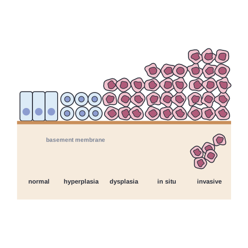
Tumour progression: normal epithelium, hyperplasia, dysplasia, carcinoma in situ and invasive cancer
This illustration explains tumour progression: normal epithelium, hyperplasia, dysplasia, carcinoma in situ and invasive cancer. Add it to cancer biology diagrams, protocol figures, slides or lessons that involve oncology. Editable SVG from the Cancer Biology Pro pack, with grouped layers you can recolor.
Format: SVG (editable vector). License: Sciicons Pro license. Courtesy: Sciicons. Part of the Cancer Biology pack.
Keywords: Cancer Biology Pack, Sciicons Pro, tumor progression, hyperplasia, dysplasia, carcinoma in situ, invasive cancer, basement membrane, cancer biology, oncology, tumor, cancer
Related cancer biology illustrations
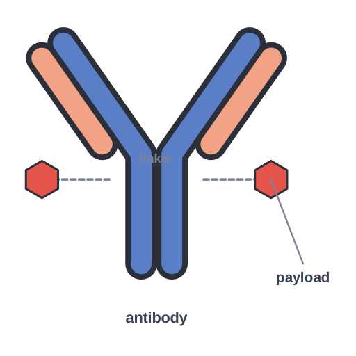 Antibody–drug conjugate (ADC) · Pro
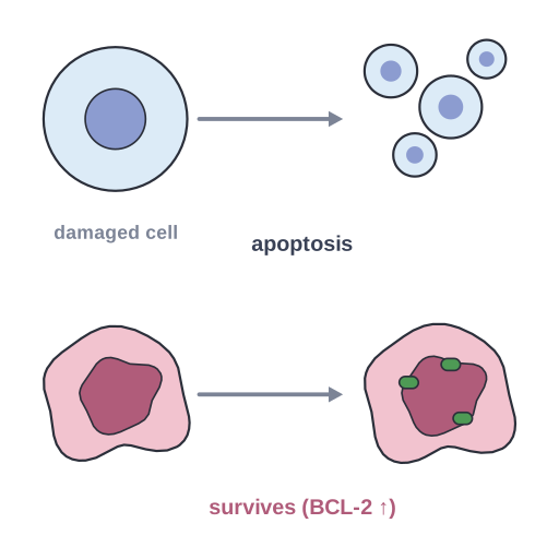 Apoptosis evasion · Pro
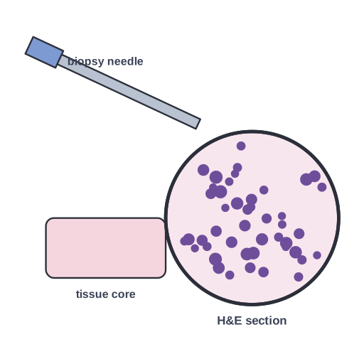 Biopsy and histology · Pro
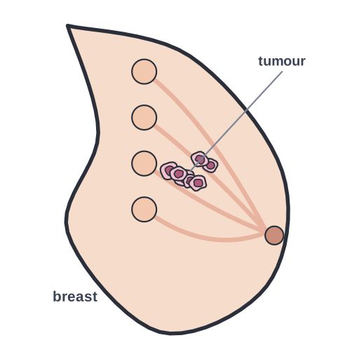 Breast cancer · Pro
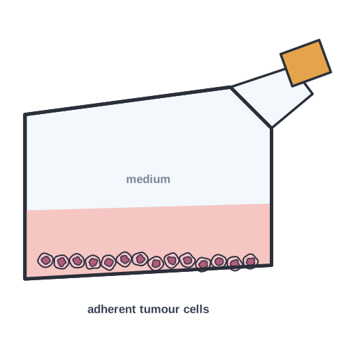 Cancer cell line grown in a culture flask with medium, as a… · Pro
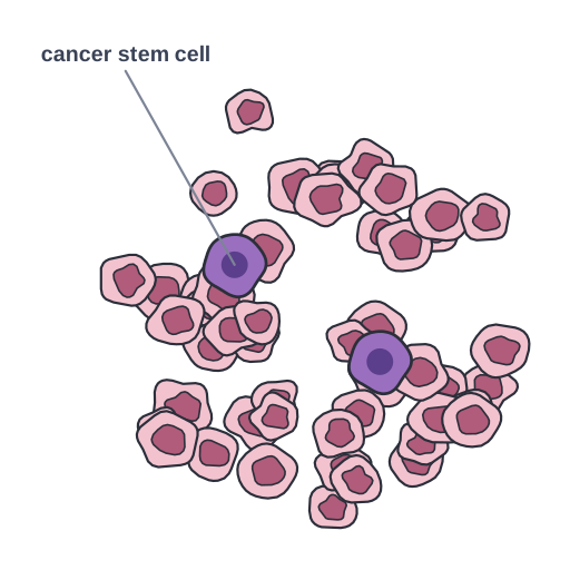 Cancer stem cells · Pro
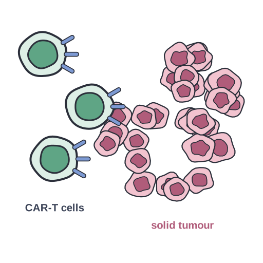 CAR-T cells infiltrating a solid tumour and binding tumour antigen · Pro
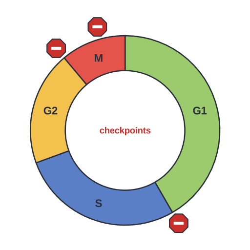 Cell cycle with G1, S, G2 and M phases and the G1/S, G2/M and… · Pro
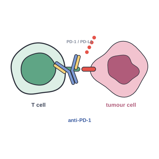 Checkpoint immunotherapy · Pro
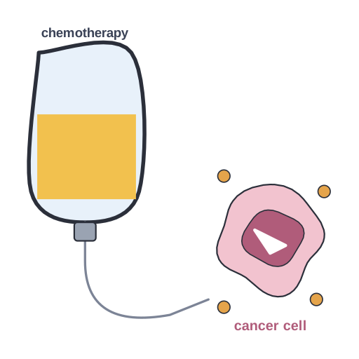 Chemotherapy · Pro
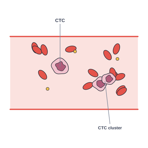 Circulating tumour cells and clusters travelling in the bloodstream… · Pro
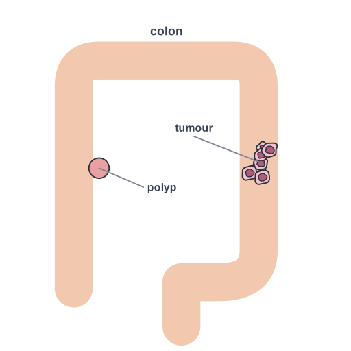 Colorectal cancer · Pro