Ear anatomy
♛ PRO Part of the Human Organs pack · 40 icons
- Every Pro pack, plus future ones
- Icon editor and recolour
- Molecules, Proteins, Gel Builder and Well Plate exports
- Use in papers, talks and commercial work
$49 one-time (₹2,999 in India) · lifetime access · no subscription
Pro keeps the free library free.
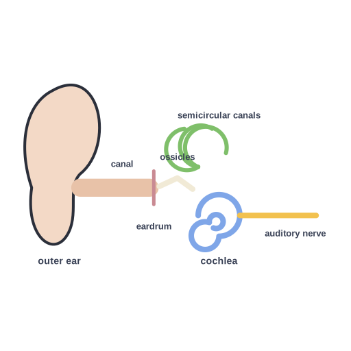
Ear anatomy: outer ear and canal, eardrum, middle-ear ossicles, cochlea, semicircular canals and auditory nerve
This illustration explains ear anatomy: outer ear and canal, eardrum, middle-ear ossicles, cochlea, semicircular canals and auditory nerve. Handy for explaining human organs in anatomy and physiology presentations, theses, posters and course notes. Editable SVG from the Human Organs Pro pack, with grouped layers you can recolor.
Format: SVG (editable vector). License: Sciicons Pro license. Courtesy: Sciicons. Part of the Human Organs pack.
Keywords: Human Organs Pack, Sciicons Pro, ear, anatomy, human organ, anatomy, human organs, anatomy, physiology
Related human physiology illustrations
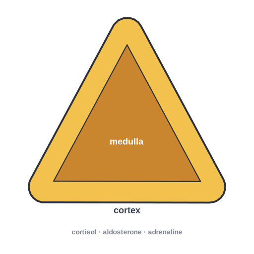 Adrenal gland section · Pro
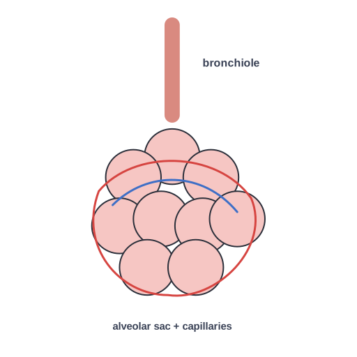 Alveoli · Pro
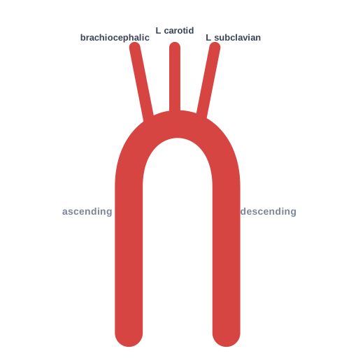 Aorta and its arch branches · Pro
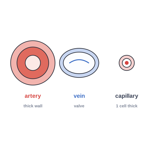 Blood vessel types · Pro
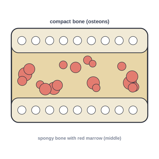 Bone section · Pro
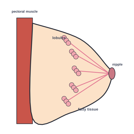 Breast anatomy (sagittal) · Pro
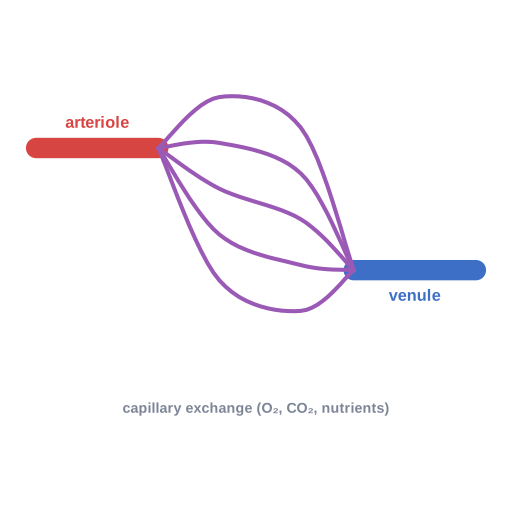 Capillary bed connecting an arteriole and a venule, where exchange… · Pro
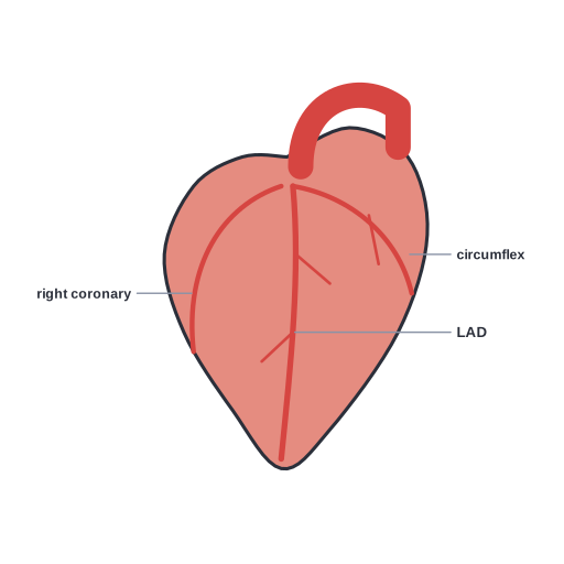 Coronary arteries supplying the heart muscle · Pro
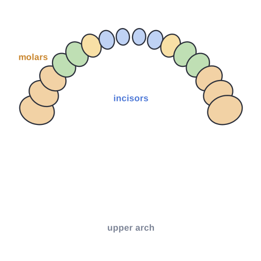 Adult dental arch · Pro
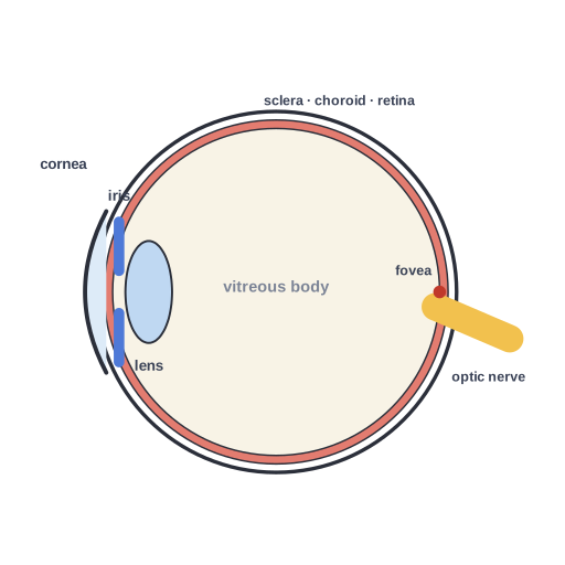 Eye cross-section · Pro
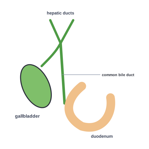 Gallbladder and biliary tree · Pro
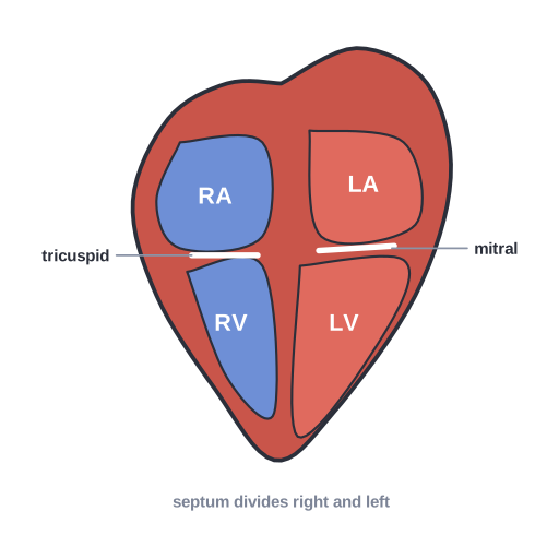 Heart cross-section · Pro