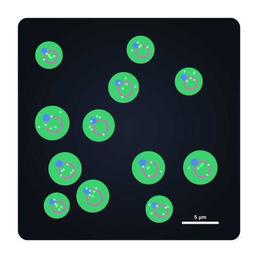
Fluorescence micrograph of yeast
♛ PRO Part of the Yeast pack · 25 icons
- Every Pro pack, plus future ones
- Icon editor and recolour
- Molecules, Proteins, Gel Builder and Well Plate exports
- Use in papers, talks and commercial work
$49 one-time (₹2,999 in India) · lifetime access · no subscription
Pro keeps the free library free.
Fluorescence micrograph of yeast: GFP-tagged protein (green) with nuclei (blue) and vacuole membranes (magenta, FM4-64 style) on a dark field
Fluorescence micrograph of yeast illustration showing GFP-tagged protein (green) with nuclei (blue) and vacuole membranes (magenta, FM4-64 style) on a dark field. Suited to microbiology manuscripts, grant figures, posters and teaching slides that cover saccharomyces cerevisiae. Available with Sciicons Pro in the Yeast pack as a layered, editable SVG.
Format: SVG (editable vector). License: Sciicons Pro license. Courtesy: Sciicons. Part of the Yeast pack.
Keywords: Yeast Pack, Sciicons Pro, fluorescence microscopy, GFP, micrograph, yeast, saccharomyces, saccharomyces cerevisiae, cerevisiae, budding yeast, fungi, fungus, eukaryotic microbe, microbiology
Related microbiology illustrations
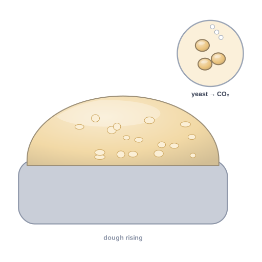 Baker’s yeast leavening bread dough · Pro
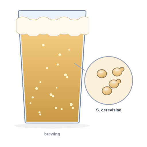 Brewing with yeast · Pro
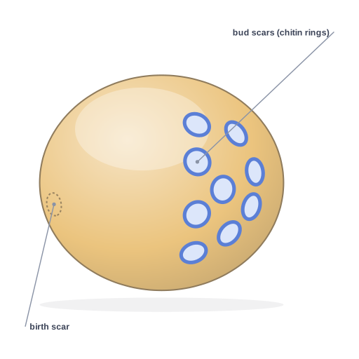 Yeast bud scars · Pro
 Candida albicans morphological switch · Pro
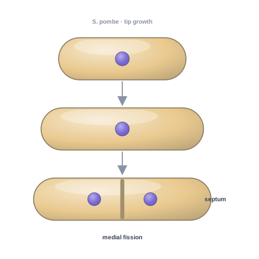 Fission yeast Schizosaccharomyces pombe · Pro
 Fission yeast (S. pombe) silhouette · Pro
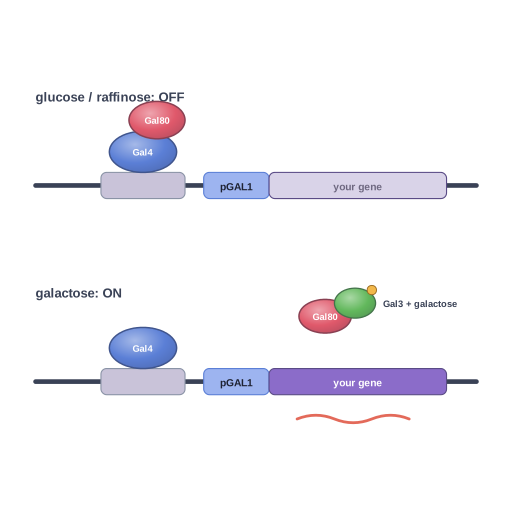 GAL promoter induction in yeast · Pro
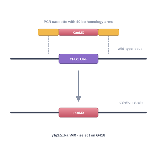 Yeast gene knockout by homologous recombination · Pro
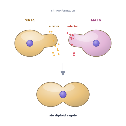 Yeast mating · Pro
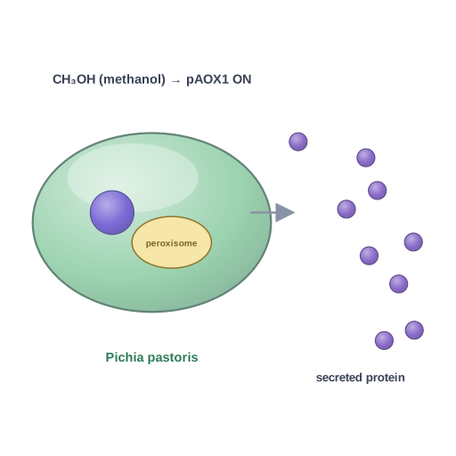 Pichia pastoris (Komagataella) expression host · Pro
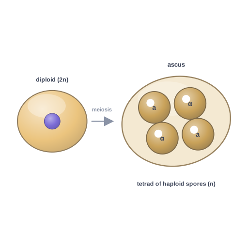 Yeast sporulation · Pro
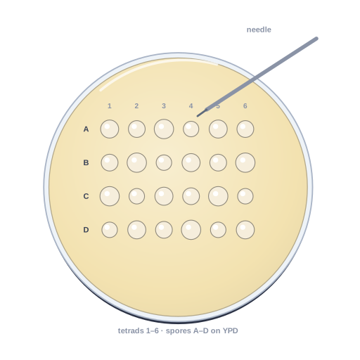 Tetrad dissection · Pro