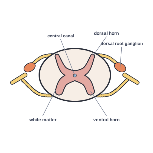
Spinal cord cross-section
♛ PRO Part of the Neuroscience pack · 47 icons
- Every Pro pack, plus future ones
- Icon editor and recolour
- Molecules, Proteins, Gel Builder and Well Plate exports
- Use in papers, talks and commercial work
$49 one-time (₹2,999 in India) · lifetime access · no subscription
Pro keeps the free library free.
Spinal cord cross-section: butterfly-shaped grey matter with dorsal and ventral horns, surrounding white matter, central canal and spinal nerve roots
This illustration explains spinal cord cross-section: butterfly-shaped grey matter with dorsal and ventral horns, surrounding white matter, central canal and spinal nerve roots. Researchers and educators can drop it into neuroscience slides, review figures or classroom handouts on neurobiology. Part of the Sciicons Neuroscience pack (Pro), delivered as an editable SVG with named layers.
Format: SVG (editable vector). License: Sciicons Pro license. Courtesy: Sciicons. Part of the Neuroscience pack.
Keywords: Neuroscience Pack, Sciicons Pro, spinal cord, cross-section, grey matter, white matter, dorsal horn, ventral horn, central canal, dorsal root ganglion, spinal nerve, neuroscience, neurobiology, brain
Related neuroscience illustrations
 Action potential · Pro
 Acute extracellular recording · Pro
 Alzheimer’s disease · Pro
 Astrocyte · Pro
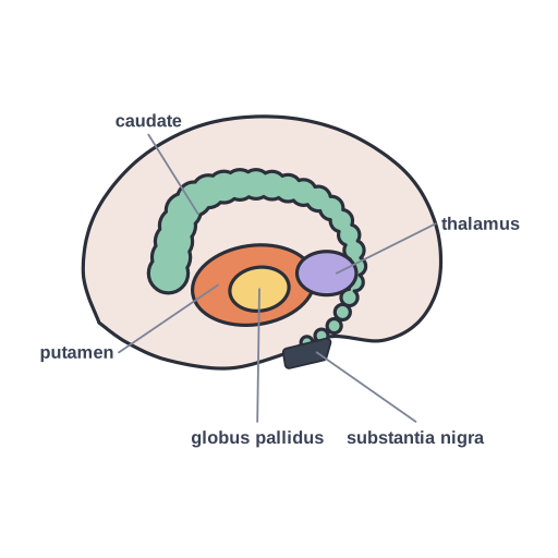 Basal ganglia · Pro
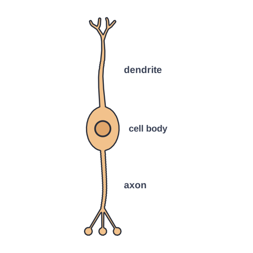 Bipolar neuron · Pro
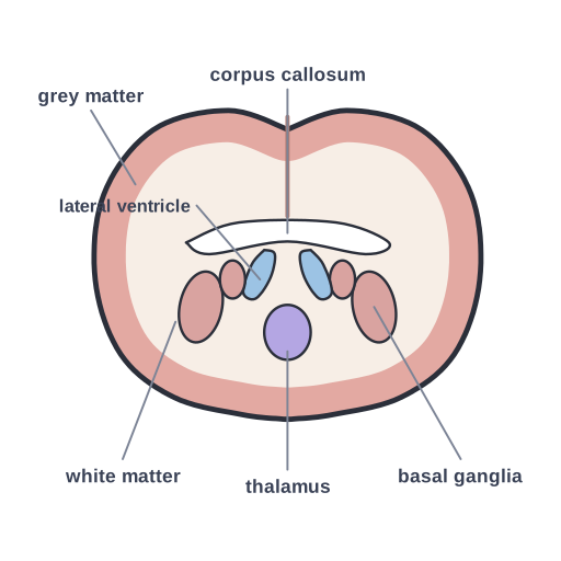 Human brain, coronal section · Pro
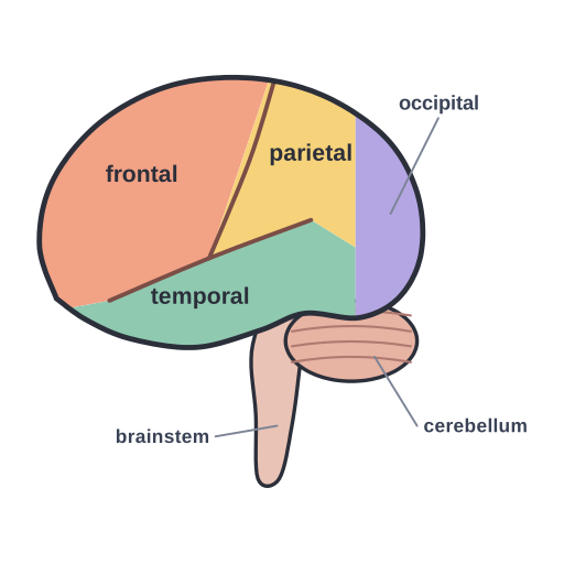 Human brain, lateral view · Pro
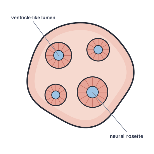 Brain organoid · Pro
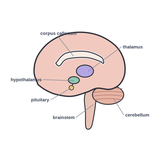 Human brain, midsagittal section · Pro
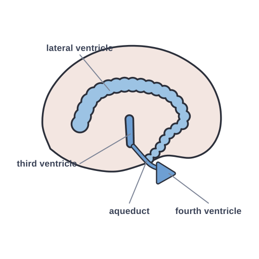 Ventricular system · Pro
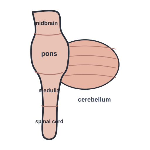 Brainstem and cerebellum · Pro