Epidermal cells
♛ PRO Part of the Human Tissues & Cells pack · 40 icons
- Every Pro pack, plus future ones
- Icon editor and recolour
- Molecules, Proteins, Gel Builder and Well Plate exports
- Use in papers, talks and commercial work
$49 one-time (₹2,999 in India) · lifetime access · no subscription
Pro keeps the free library free.
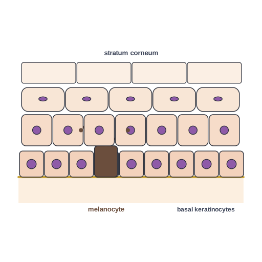
Epidermal cells: basal keratinocytes, melanocytes passing pigment, flattening keratinocytes and cornified layer
This illustration explains epidermal cells: basal keratinocytes, melanocytes passing pigment, flattening keratinocytes and cornified layer. It works well in histology figures, teaching materials and conference posters that explain human tissue. Editable SVG from the Human Tissues & Cells Pro pack, with grouped layers you can recolor.
Format: SVG (editable vector). License: Sciicons Pro license. Courtesy: Sciicons. Part of the Human Tissues & Cells pack.
Keywords: Human Tissues & Cells Pack, Sciicons Pro, epidermis, cells, histology, human tissue, cell, histology, human tissues, cell biology
Related tissues illustrations
 White adipocyte · Pro
 Adipose tissue · Pro
 Alveolar wall cells · Pro
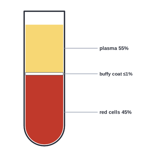 Blood composition after centrifugation · Pro
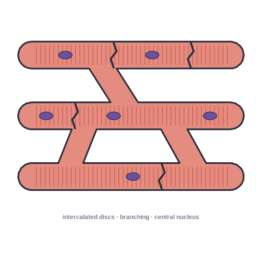 Cardiac muscle tissue · Pro
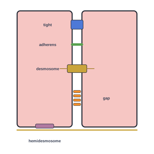 Cell junctions between two epithelial cells · Pro
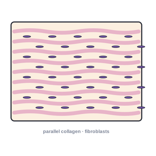 Dense regular connective tissue (tendon, ligament) · Pro
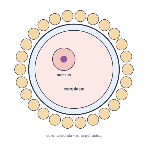 Egg cell (oocyte) · Pro
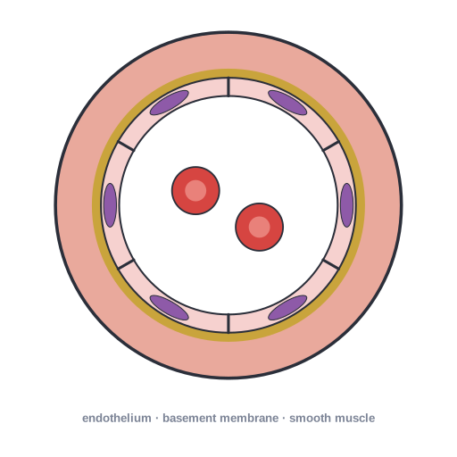 Endothelial cells lining a blood vessel lumen · Pro
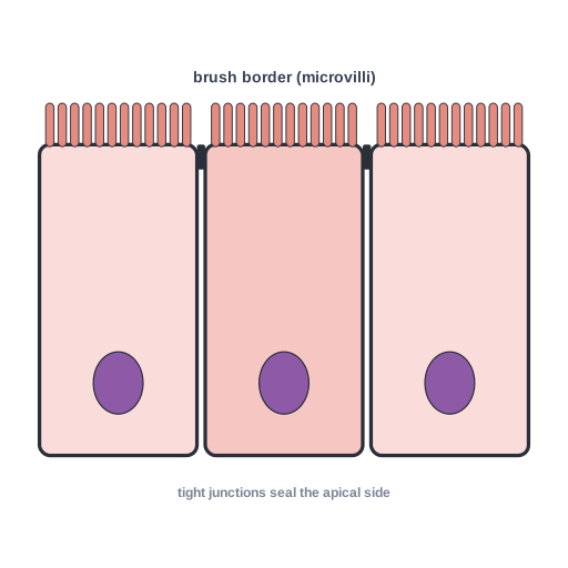 Enterocyte with apical microvilli (brush border), tight junctions… · Pro
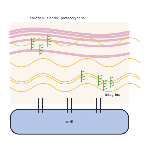 Extracellular matrix · Pro
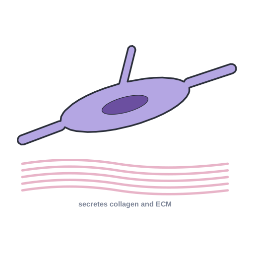 Fibroblast · Pro